Welcome to InView Imaging: The Bay Area MRI Provider
At InView Imaging, we understand that undergoing an MRI can be stressful. That’s why we are dedicated to providing the highest level of care and comfort.
We are thrilled to announce the opening of our new open MRI facility in Concord. Unlike traditional MRI machines, our state-of-the-art open MRI offers a more spacious and comfortable experience, ideal for patients who feel claustrophobic or anxious. This advanced technology ensures high-quality imaging while keeping you relaxed throughout the process.
MRI, or Magnetic Resonance Imaging, is a non-invasive procedure used to diagnose and monitor a variety of conditions. It provides detailed images of soft tissues, organs, and other internal structures, making it essential for detecting issues in the brain, spine, joints, and abdomen. MRI is also invaluable for evaluating conditions like tumors, internal bleeding, and infections.
Our team of compassionate and experienced radiologists and staff is here to provide personalized care tailored to your specific needs. From the moment you arrive, you will be greeted by our friendly staff, ready to guide you through every step and answer any questions you may have.
In addition to our Concord location, InView Imaging proudly serves the community with facilities in Fremont, Lafayette, Antioch, and San Ramon. We accept most insurance plans, making our services accessible and stress-free. Trust us to provide exceptional radiology services with a personal touch. Schedule your appointment at any of our convenient locations today and experience the InView difference.
Ever wondered how medical professionals get detailed insights into the human body? The answer lies in one powerful tool: MRI. From detecting ailments to guiding treatments, MRI technology, with its scanner and magnetic field, revolutionizes healthcare in clinical practice for the patient. But what exactly is MRI, and how does it work its magic by using a magnetic field and scanner to create images? Let’s delve into the fascinating world of magnetic resonance imaging, mri scanners, mri exam, mri technician, and uncover its secrets. Ready to unlock the mysteries within your body with just an MRI scan? Stay tuned as we explore the incredible capabilities and benefits of MRI technology, including the magnetic field, contrast agents, and images, that have transformed modern medicine.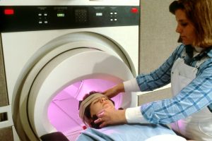
MRI Essentials
Importance Explained
MRI, using a magnetic field and contrast agents, plays a crucial role in diagnosing various medical conditions such as brain tumors and spinal cord injuries. It helps healthcare providers detect abnormalities early, enabling prompt treatment interventions. The non-invasive nature of MRI makes it preferred over invasive procedures like exploratory surgery.
Key Processes
The principles of magnetic resonance imaging involve utilizing powerful magnets and radio waves to create detailed images of the body’s internal structures. To create an MRI image, the patient lies inside a large tube-like machine where radio waves generate signals from the body’s tissues and magnetic field. Contrast agents are often used during MRI scans to enhance visibility of specific areas or organs, aiding in accurate diagnosis.
Understanding Results
Interpreting MRI reports requires understanding the difference between normal and abnormal findings. Normal results indicate healthy tissue appearances, while abnormal results may show signs of injury or disease. Consulting with a radiologist is essential for result analysis as they are trained to interpret complex imaging studies accurately.
MRI Diagnosis
MRI Significance
MRI plays a crucial role in modern medicine by providing detailed images of the brain with the help of contrast agents, aiding in accurate diagnosis. This imaging technique has revolutionized diagnostic procedures, allowing doctors to detect tumors and abnormalities with precision. The significance of MRI lies in its ability to offer non-invasive insights into the brain’s structure and function.
With the advancement of technology, MRI scans have become instrumental in enabling personalized healthcare for patients. By capturing high-resolution images, MRI helps doctors tailor treatment plans based on individual needs. The detailed information obtained from an MRI scan assists medical professionals in making informed decisions regarding surgery or other interventions.
Procedure Overview
When undergoing an MRI scan, patients are required to lie still inside a large tube-like machine that generates magnetic fields and radio waves to create detailed images of the brain. The process is painless and does not involve any radiation exposure, making it a safe diagnostic tool. Patients may experience slight discomfort due to the confined space during the procedure.
An average MRI procedure typically lasts between 30 minutes to an hour, depending on the complexity of the scan required. It is essential for individuals undergoing an MRI to remain motionless throughout the process to ensure clear and accurate imaging results. Any movement can distort the images and affect the diagnostic quality.
During an MRI scan, patients are instructed to follow specific guidelines such as removing metallic objects like jewelry or hearing aids that can interfere with the magnetic field. Following these instructions is crucial for obtaining precise images needed for an accurate diagnosis. Individuals with pacemakers or metal implants must inform their healthcare provider before undergoing an MRI scan.
MRI for Various Organs
Brain and Spine
MRI scans of the brain and spine are commonly performed to detect tumors, injuries, or abnormalities. The non-invasive nature of MRI makes it ideal for evaluating neurological conditions without surgery. By providing detailed images, MRI assists in diagnosing conditions like multiple sclerosis or spinal cord injuries accurately.
Heart and Vessels
In assessing heart health, MRI plays a crucial role by producing high-resolution images of the heart and blood vessels. Compared to other imaging techniques, cardiac MRI offers superior soft tissue contrast for precise diagnosis. Early detection of heart diseases through MRI enables timely intervention and treatment.

Internal Organs
The versatility of MRI extends to imaging internal organs such as the liver, kidneys, and pancreas with exceptional clarity. Evaluating organ function through dynamic imaging sequences helps identify issues like cirrhosis or renal abnormalities effectively. Detecting tumors and other anomalies in internal organs is another key strength of MRI technology.
Bones and Joints
For diagnosing bone fractures or joint disorders, MRI stands out due to its ability to capture detailed images with high resolution. It excels in evaluating musculoskeletal injuries by precisely visualizing soft tissues like ligaments or cartilage. The intricate visualization capability of MRI aids healthcare providers in accurate diagnosis and treatment planning.
Specialized MRI Exams
Breast MRI
Breast MRI plays a crucial role in detecting breast cancer, offering detailed images that aid in early detection. This specialized MRI exam is recommended for individuals with a high risk of breast cancer due to genetic factors or family history. The sensitivity of MRI in detecting breast abnormalities surpasses that of traditional mammograms.
When considering the effectiveness of MRI exams, it’s essential to understand the benefits they offer in specific scenarios like breast cancer screening. A breast MRI is often recommended for women with dense breast tissue or those who have previously undergone radiation therapy. The detailed images produced by this medical imaging technique help identify abnormalities that may be missed by other diagnostic tools.

The superior sensitivity of a breast MRI compared to mammograms makes it a valuable tool for early detection and monitoring of breast conditions. With its ability to detect smaller lesions and assess the extent of disease spread, an MRI exam provides critical information for healthcare providers when making treatment decisions.
Preparing for MRI
Risks Identified
MRI scans are generally safe; however, potential risks may include discomfort from lying still, loud noises, and potential reactions to contrast agents. To minimize risks during an MRI scan, safety measures such as proper screening for metal objects are crucial. In rare instances, patients may experience adverse reactions to MRI contrast agents like nausea or allergic reactions.
Preparation Tips
When preparing for an MRI scan, ensure you inform the healthcare provider about any metal implants or devices in your body. Follow dietary restrictions, which typically involve avoiding food a few hours before the procedure. It is essential to understand that certain metals can interfere with the magnetic field of the MRI machine.
During the MRI Scan
Expectations Set
When undergoing an MRI scan, it’s crucial to manage your expectations for a smooth experience. The mri scanner generates loud noises, akin to thumping or knocking sounds. Claustrophobia concerns are common due to the tight space inside the machine.
To ensure accurate results, it is vital to remain perfectly still during the mri scans. Even slight movements can blur the images and affect diagnosis accuracy significantly.
Procedure Steps
The process of undergoing an MRI scan involves several critical steps. Firstly, you will be asked to remove all metal objects like jewelry or clothing with metal fastenings.
Next, you’ll lie on a padded table that slides into the mri scanner – a large tube surrounded by powerful magnets and radio waves. The machine creates a strong magnetic field that aligns hydrogen atoms in your body.
The radio waves then disrupt this alignment temporarily, producing signals used to create detailed cross-sectional images of your body’s internal structures. Depending on the type of scan (T1 or T2), different contrasts may be achieved for enhanced visualization.
Throughout the procedure, an MRI technologist will guide you and ensure your comfort and safety in the scanning environment. Their expertise plays a crucial role in obtaining high-quality images while addressing any patient concerns promptly.
After the MRI Scan
Post-Procedure Care
After completing an MRI scan, it is crucial to follow specific guidelines for post-procedure care. Firstly, ensure to drink plenty of water to help flush out the contrast dye used during the scan. Hydration aids in a quicker recovery process.
You can generally resume normal activities immediately after an MRI scan. However, it is advisable to avoid any strenuous physical activities for a few hours post-scan. It’s essential to take note of any specific instructions provided by the healthcare provider.
Follow-Up Actions
Once you receive your MRI results, the next step involves understanding and discussing them with your healthcare provider. It’s vital to schedule a follow-up appointment to go over the findings in detail and determine any necessary treatment plans or further tests.
Regular follow-up appointments with your healthcare provider are crucial after an MRI scan. These appointments allow for continuous monitoring of your health condition and treatment progress based on the initial findings from the MRI results. The information obtained from these scans plays a significant role in guiding future medical decisions and interventions.
Interpreting MRI Results
Results Breakdown
An MRI report typically includes detailed information on various aspects of the scanned area. It breaks down components like T1-weighted images, which highlight anatomy, and T2-weighted images, showing fluid accumulation. The terminology may mention lesions (abnormal tissue changes) or enhancement (increased signal due to blood flow).
Understanding MRI results involves grasping the significance of different findings. For instance, detecting a tumor can indicate the need for further evaluation or treatment. Conversely, identifying a herniated disc might explain back pain symptoms. Each finding in an MRI report provides crucial insights into potential health conditions.
Next Steps
Upon receiving MRI results, the next step is consulting with a healthcare professional for thorough interpretation. Based on the findings, treatment options vary; for example, surgery may be necessary for conditions like tumors or severe injuries. In contrast, conservative approaches such as physical therapy could suffice for less critical issues.
Discussing MRI results with a healthcare provider is essential to develop an appropriate treatment plan tailored to individual needs. This ensures that patients receive personalized care aligned with their specific diagnosis and medical history. By collaborating with medical experts, individuals can make informed decisions regarding their health and well-being.
MRI Safety and Innovations
Technological Advances
MRI imaging has seen significant advancements in recent years, with the introduction of 3T and 7T MRI machines. These higher-field-strength scanners provide enhanced image resolution, allowing for better visualization of tissues and organs. The development of functional MRI (fMRI) enables the mapping of brain activity by detecting changes in blood flow.
Advanced MRI techniques offer numerous benefits, including improved diagnostic accuracy and early detection of abnormalities. For example, diffusion-weighted imaging (DWI) is highly sensitive to tissue changes, making it valuable in diagnosing strokes promptly. Spectroscopy provides insights into tissue metabolism, aiding in cancer diagnosis and treatment planning.
Technological innovations have revolutionized MRI by enhancing both accuracy and efficiency. The integration of artificial intelligence (AI) algorithms helps in image reconstruction, reducing scan times while maintaining high image quality. Moreover, real-time MRI guidance during procedures such as biopsies ensures precision and safety for patients.
Safety Measures
During an MRI scan, strict safety protocols are followed to ensure patient well-being. Patients are required to remove all metallic objects before entering the scanning room to prevent injuries from magnetic forces. Furthermore, continuous monitoring by trained technologists is crucial to address any potential issues promptly.
Screening for contraindications before an MRI procedure is vital to prevent adverse events. Patients with certain implants or medical devices may not be suitable candidates for an MRI due to safety concerns related to magnetic fields. Proper screening helps identify individuals at risk and ensures their safety during the scan.
Trained professionals play a key role in maintaining MRI safety throughout the imaging process. Radiologic technologists are responsible for operating the scanner correctly and monitoring patients for any signs of distress or discomfort during the scan. Their expertise ensures that patients receive high-quality imaging while minimizing risks associated with the procedure.
Closing Thoughts
You’ve now gained a comprehensive understanding of MRI essentials, its role in diagnosing brain tumors and various organ conditions, specialized MRI exams, preparation, scan process, result interpretation, safety measures, and innovations. Remember, MRI is a powerful tool that aids in accurate diagnoses and treatment planning. Stay informed about the latest advancements to ensure you receive the best care possible.
As you navigate the world of medical imaging, always prioritize your health and well-being. If you have any concerns or require further information about MRI procedures, don’t hesitate to consult with healthcare professionals. Your proactive approach to healthcare empowers you to make informed decisions for your overall wellness.
Frequently Asked Questions
What are the essential aspects of MRI?
MRI is a non-invasive imaging technique that provides detailed images of internal body structures. It is crucial for diagnosing various conditions without radiation exposure, making it safe and effective for patients of all ages.
How does an MRI help in diagnosing brain tumors?
MRI plays a vital role in the diagnosis of brain tumors by providing high-resolution images that allow healthcare professionals to visualize the size, location, and characteristics of the tumor. This information is critical for treatment planning and monitoring.
Which organs can be examined using MRI?
MRI can be used to examine various organs in the body, including the brain, spine, heart, liver, kidneys, and reproductive organs. It offers detailed insights into organ structure and function without invasive procedures or exposure to harmful radiation.
What should I do to prepare for an MRI scan?
Before an MRI scan, you may need to remove any metal objects and inform the technologist about any medical conditions or implants you have. It’s essential to follow specific instructions provided by your healthcare provider for a successful scan.
What happens during an MRI scan?
During an MRI scan, you will lie on a table that slides into the scanner. You need to stay still while the machine captures detailed images of your body parts. The process is painless and typically takes around 15-60 minutes depending on the type of exam.
How soon can I expect my MRI results after the scan?
After completing your MRI scan, a radiologist will interpret the images and generate a report detailing their findings. Depending on your healthcare provider’s practice, you may receive your results within a few days to discuss further steps if needed.
Are there any safety measures or innovations in MRI technology?
MRI technology continues to evolve with enhanced safety features such as reducing noise levels during scans and improving image quality for better diagnostics. Innovations like open-bore systems provide more comfort for patients while maintaining high accuracy in imaging results.