Mammography
2D Digital Mammography and 3D Tomographic Mammography
Schedule your mammogram today with our compassionate, efficient staff.
Click the Live Help feature below to find the next available appointment.
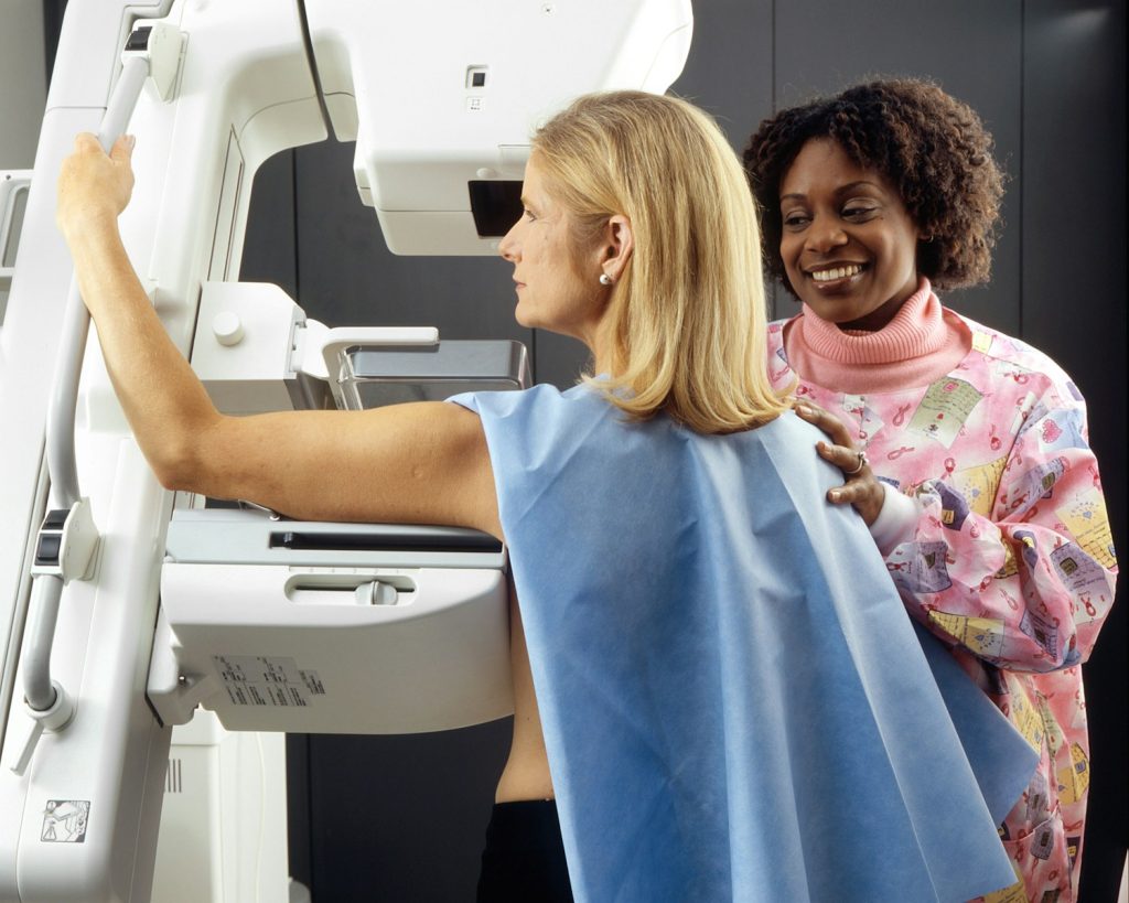
A mammogram uses X-ray radiation to create images of the breast tissue.
Offered at these locations:
2D Digital in Oakland, Lafayette, Antioch, and Fremont.
3D Tomographic in Lafayette and Fremont.
Cash Price:
$200 for Screening
$150 for Diagnostic Unilateral
$350 for Diagnostic Bilateral
Mammograms have been a vital tool in detecting breast cancer for decades. With advancements in technology, the transition from 2D to 3D mammograms (dbt) has revolutionized early detection capabilities. The traditional 2D mammogram provides a flat image, while the newer 3D DBT version offers a clearer, more detailed view of breast tissue with a lower radiation dose. This enhanced imaging, including standard 2d mammogram images and dbt, can lead to earlier detection of abnormalities and potentially reduce false positives, providing women with greater peace of mind during screenings.
The evolution from 2D to 3D mammograms marks a significant milestone in improving breast cancer screening accuracy and effectiveness. Understanding the differences between these two technologies is crucial for women’s health and well-being.
Mammogram Basics
2D vs 3D
2D mammograms provide flat images and a higher dose, while 3D mammograms (dbt) offer a more detailed three-dimensional view. The 3D technology captures multiple images from various angles, providing a clearer picture of breast tissue. This advancement enhances the ability to detect abnormalities that may be hidden in traditional 2D scans, mammogram images, dbt, and dose.
The benefits of 3D imaging, including standard 2D mammogram images, dose, and dbt, include improved accuracy in detecting breast cancer, especially in women with dense breast tissue. Compared to standard 2D mammograms, 3D (dbt) mammograms reduce the need for additional screenings due to their higher sensitivity. The impact on detection accuracy is significant, as 3D imaging can detect smaller tumors earlier, leading to better treatment outcomes.
Image Collection
During a mammogram, specialized equipment, including digital breast tomosynthesis (DBT), captures X-ray images of the breasts from different angles. High-quality images are crucial for accurate interpretation by radiologists. Advancements in image collection techniques have led to clearer and more precise mammogram images, aiding in early detection of breast cancer.
The process involves positioning the breast between two plates for compression while images are taken. High-resolution digital systems ensure detailed imaging necessary for identifying abnormalities like tumors or calcifications. Improved image quality allows for better visualization of breast tissue structures, enhancing diagnostic capabilities.
Compression Consistency
Consistent compression during mammograms is essential for optimal image quality and reliable results. Proper compression spreads out breast tissue evenly, reducing overlapping structures that could obscure abnormalities. Inconsistent compression can lead to distorted images and potential missed detections.
Compression plays a vital role in maintaining uniform thickness across the entire breast, ensuring an even distribution of X-rays during imaging. Radiologic technologists follow specific guidelines to apply adequate pressure without causing discomfort or compromising image quality. Maintaining consistent compression levels throughout the procedure is critical for accurate interpretation by healthcare providers.
Image Clarity
2D Limitations
2D mammograms, while effective, have limitations in detecting abnormalities due to their flat imaging nature. In scenarios where abnormalities overlap or hide behind dense breast tissue, 2D imaging may miss crucial details. This can lead to false negatives and delayed diagnosis, emphasizing the importance of additional screening methods.
The flat dimensional picture provided by 2D mammograms sometimes fails to offer a comprehensive view of the breast tissue. For women with dense breasts or those prone to developing masses in overlapping areas, envision imaging through 2D scans may not be sufficient. This underlines the necessity for supplementary screenings like ultrasound or MRI for a more thorough examination.
There is a growing recognition within the medical community regarding the need for supplemental screening methods alongside 2D mammograms. While 2D imaging remains valuable as an initial screening tool, its limitations necessitate additional measures like insurance-covered 3D mammograms for improved accuracy and early detection.
3D Advantages
One of the key advantages of 3D mammograms over traditional 2D imaging is their ability to provide a more detailed and layered view of breast tissue. By capturing images from multiple angles, 3-dimensional image technology enhances detection rates by offering radiologists a clearer picture with reduced chances of missing abnormalities.
The advanced technology utilized in 3D mammography significantly reduces false positives compared to traditional 2D scans. This means fewer instances where benign findings are mistaken for cancerous growths, leading to unnecessary anxiety and follow-up tests for patients. The improved accuracy offered by clear plastic plate-based envision imaging helps streamline the diagnostic process and minimizes unnecessary interventions.
With enhanced clarity and precision in detecting abnormalities, 3-dimensional pictures, generated through cutting-edge technology used in 3D mammography machines, provide healthcare providers with detailed insights into breast health. The sharper images facilitate early intervention strategies and treatment plans tailored to individual patient needs.
Understanding Breast Density
Density Importance
Breast density plays a crucial role in mammogram screenings as it refers to the amount of glandular and fibrous tissue compared to fatty tissue. Higher breast density can make it challenging to detect abnormalities on mammograms, increasing the risk of missed diagnoses. In contrast, lower breast density allows for clearer imaging and easier detection of potential issues.
Breast density significantly impacts their effectiveness. 2D mammograms may struggle to differentiate between dense tissue and possible tumors due to overlapping structures, potentially leading to false positives or negatives. On the other hand, 3D mammography, also known as tomosynthesis, provides clearer images by capturing multiple angles of the breast, enhancing visibility even in dense breasts.
The challenges associated with dense breast tissue extend beyond screening accuracy. Women with higher breast density are at an increased risk of developing breast cancer themselves, making early detection through effective screenings vital. Dense tissue can mask abnormalities on traditional 2D scans, highlighting the necessity for advanced imaging technologies like 3D mammography.
Impact on Detection
Mammogram technology is instrumental in detecting early signs of breast cancer, offering a crucial advantage in successful treatment outcomes. The transition from traditional 2D mammograms to 3D imaging has revolutionized early detection capabilities by providing detailed cross-sectional views that help radiologists identify small abnormalities that might have been overlooked previously.
Incorporating 3D imaging into routine screenings has shown significant improvements in detecting invasive cancers and reducing unnecessary callbacks for additional tests caused by false alarms from conventional methods. The ability of 3D mammography to capture multiple images at different angles enhances radiologists’ ability to pinpoint suspicious areas accurately.
Regular screenings are essential for improving detection outcomes as they enable healthcare providers to monitor changes in breast tissue over time effectively. By undergoing annual mammograms, women can increase their chances of detecting any anomalies promptly, allowing for timely intervention and improved prognosis if any issues arise.
Tomosynthesis is a three-dimensional mammogram that uses X-rays to obtain sectional images of the breast, which are then reconstructed into a 3D volume. Tomosynthesis is performed by moving the X-ray tube on a circular arch and making a series of low-dose exposures, from different angles, in a few seconds.
Cancer Detection
2D Challenges
2D mammograms, while effective, come with challenges. One limitation is the inability to differentiate overlapping breast tissue, potentially leading to missed abnormalities. Factors like breast density can also impact the accuracy of 2D imaging results. As breast density increases, detection sensitivity decreases, making it harder to identify potential cancers.
Complementary screening methods are crucial when using 2D technology. Supplementing with ultrasound or MRI scans can help capture details that may be obscured in a 2D image. These additional screenings provide a more comprehensive view of the breast, especially for women with dense breast tissue. It’s essential to consider these challenges and explore alternative screening options for improved cancer detection.
3D Efficacy
3D mammograms, also known as tomosynthesis, offer enhanced efficacy in detecting breast abnormalities compared to traditional 2D imaging. Studies have shown that 3D mammography detects more cancers than 2D alone and reduces the need for additional tests due to false positives. The three-dimensional images generated by this technology provide radiologists with a clearer view of the breast tissue, improving diagnostic accuracy.
Statistics support the effectiveness of 3D imaging in early cancer detection. Research indicates that 3D mammograms increase cancer detection rates by up to 40%, particularly in women with dense breasts where tumors may be harder to spot on a conventional mammogram. The ability of 3D technology to pinpoint abnormalities earlier can lead to timely interventions and better treatment outcomes for patients.
Advantages of 3D Mammograms
Reduced False Alarms
3D mammograms minimize false alarms by providing a clearer view of breast tissue layers. This advanced technology reduces unnecessary biopsies and the stress associated with false positives. Patients often face anxiety and emotional distress when called back for further testing due to inaccurate results. The accuracy of 3D mammograms plays a crucial role in alleviating these concerns, ensuring peace of mind for patients.
Improved Detection Rates
Compared to traditional 2D imaging, 3D mammograms boast enhanced detection rates. By capturing multiple images from various angles, they can detect abnormalities that may be missed in 2D scans. Early detection is key in improving treatment outcomes for breast cancer patients. Studies have shown that 3D mammography significantly increases the detection of invasive cancers while reducing the need for additional imaging tests.
In one case study, a woman underwent both 2D and 3D mammograms during her routine screening. While the 2D scan showed no signs of cancer, the subsequent 3D scan revealed a small tumor that was successfully treated at an early stage. This example highlights how the improved detection capabilities of 3D mammograms can potentially save lives by detecting cancer earlier than traditional methods.
Target Audience for 3D Mammograms
High-Risk Individuals
Radiologists play a crucial role in recommending specialized screening for high-risk individuals. These screenings are essential for detecting potential breast cancer at early stages. Genetic factors and family history significantly impact an individual’s risk level, emphasizing the need for tailored screening approaches. Personalized screening plans are vital to ensure high-risk patients receive appropriate and timely interventions.
Dense Breast Tissue
Screening individuals with dense breast tissue poses unique challenges due to the difficulty in detecting abnormalities on mammograms. The accuracy of mammograms is often compromised by dense breasts, leading to lower detection rates. Consequently, women with dense breasts may benefit from supplemental screening methods, such as ultrasound or MRI scans, to enhance early detection and improve overall outcomes.
American Cancer Society Recommendations
Screening Guidelines
The American Cancer Society recommends regular mammograms for women starting at age 40, with yearly screenings thereafter. For those with a higher risk due to family history or genetic mutations, screening might start earlier. Follow-up exams are crucial if abnormalities are detected during the initial screening.
Controversies exist regarding the frequency of screenings and the ideal age to begin. Some experts argue that annual mammograms starting at age 40 save more lives, while others suggest biennial screenings from age 50 onwards. These debates highlight the need for personalized decisions based on individual risk factors.
2D vs 3D Considerations
When comparing 2D and 3D mammograms, several factors come into play. While traditional 2D imaging is widely accessible and covered by most insurance plans, 3D mammography offers improved accuracy in detecting breast cancer by providing clearer images of overlapping tissues.
Cost can be a significant consideration when opting for either type of mammogram. Although 3D mammograms may cost more, their ability to detect cancers earlier can potentially reduce treatment costs in the long run. It’s essential for individuals to weigh this financial aspect against the added benefits of enhanced detection rates.
In making an informed decision between 2D and 3D imaging, availability also plays a crucial role. While many healthcare facilities now offer both options, some centers may have limited access to 3D technology due to its higher cost and maintenance requirements. Patients should inquire about this aspect when scheduling their appointments.
Location Influence on Mammograms
Accessibility Issues
Accessing mammogram screenings can be challenging for many individuals, especially in underserved communities. Disparities exist in the availability of mammogram technology between different healthcare settings. While some facilities offer 3D mammograms, others may only provide 2D imaging services. These discrepancies contribute to unequal access to advanced screening options.
In response to these challenges, various initiatives and programs have been established to improve mammogram accessibility for underserved populations. Mobile mammography units, community outreach programs, and partnerships with local organizations aim to bring screenings closer to those who face barriers due to location or financial constraints.
Technology Availability
The availability of 3D mammogram technology varies across healthcare facilities. Larger hospitals and specialized centers are more likely to offer 3D mammograms, while smaller clinics may rely solely on traditional 2D imaging methods. This discrepancy in technology adoption impacts the quality of care and early detection outcomes for patients.
Trends indicate a gradual shift towards the integration of 3D imaging technologies in routine breast cancer screenings. Healthcare providers recognize the benefits of 3D mammograms in detecting abnormalities with greater accuracy, leading to improved diagnostic results and reduced false positives. However, cost considerations and infrastructure requirements pose challenges to the widespread implementation of this advanced technology.
Choosing the Right Mammogram
Personal Health History
Considering personal health history is crucial in determining the most suitable mammogram screening approach. Past findings and treatments significantly influence future screening recommendations. For instance, a history of breast cancer may prompt more frequent screenings or additional imaging tests.
Patient-doctor communication plays a pivotal role in developing personalized screening plans. Openly discussing previous mammogram results, family history of breast cancer, or any concerns can help tailor screening recommendations to individual needs. This collaborative approach ensures that screenings are effective and relevant.
In some cases, specific health conditions or genetic factors may necessitate early or specialized screenings. Patients should proactively share such information with their healthcare providers to ensure appropriate screening protocols are followed.
Doctor’s Advice
Seeking professional advice from healthcare providers is essential for making informed decisions about mammogram screenings. Doctors play a key role in guiding patients through the complexities of screening options and recommendations based on individual risk factors.
Healthcare providers offer valuable insights into the benefits and limitations of different mammogram technologies, such as 2D and 3D imaging. Their expertise helps patients understand the implications of various screening methods and choose the most suitable option for their needs.
Effective communication with doctors regarding mammogram concerns is vital for ensuring a smooth screening process. Patients should feel empowered to ask questions, express any anxieties they may have, and actively participate in decision-making regarding their breast health.
Final Remarks
You’ve delved into the world of 2D and 3D mammograms, understanding their differences, benefits, and impact on cancer detection. With clearer images and improved accuracy, 3D mammograms offer a promising advancement in breast cancer screening, especially for those with dense breast tissue. The American Cancer Society’s guidelines and the influence of location on mammogram availability have also been highlighted, aiding you in making informed decisions about your health.
As you navigate the realm of mammograms, remember that early detection is key in the fight against breast cancer. Stay proactive about your health, follow recommended screening guidelines, and consult with healthcare professionals to determine the best approach for you. Your commitment to regular screenings can make a significant difference in catching any potential issues early. Take charge of your health today!
Frequently Asked Questions
What are the differences between 2D and 3D mammograms?
2D mammograms provide two-dimensional images of the breast, while 3D mammograms offer three-dimensional images. The latter allows for clearer visualization of breast tissue, aiding in the detection of abnormalities that may be hidden in traditional 2D scans.
Who should consider getting a 3D mammogram?
Women with dense breast tissue or those at higher risk for breast cancer may benefit from 3D mammograms. It provides better clarity and accuracy in detecting abnormalities, especially in women with dense breasts where issues can be harder to spot on a traditional 2D scan.
Are there any disadvantages to opting for a 3D mammogram over a 2D one, in terms of radiation dose and the dimensional image quality for the radiologist?
While not necessarily disadvantages, some considerations include slightly higher radiation exposure and potentially higher costs associated with 3D mammography. However, the improved accuracy and lower recall rates often outweigh these factors for many individuals seeking comprehensive breast screenings.
How does breast density impact the effectiveness of mammograms?
Breast density refers to the amount of glandular and connective tissue compared to fatty tissue in the breasts. Higher breast density can make it challenging to detect abnormalities on standard mammograms as both tumors and dense tissue appear white on imaging. Therefore, additional screening methods like 3D mammography are recommended for women with dense breasts.
How do location factors influence access to different types of mammograms?
The availability of advanced technologies like 3D mammography may vary based on geographical location or healthcare facilities’ resources. Urban areas or specialized medical centers might offer more options such as both traditional 2D and advanced 3D screenings compared to rural settings where access could be limited primarily to standard procedures.