Key Takeaways
-
Advanced high-field MRI systems, by definition, operate at 3 Tesla and above. They provide clearer and crisper images than conventional MRI, enabling earlier and more precise diagnoses for American patients.
-
Advanced High-Field MRI These powerful, high-resolution scanners are revolutionizing care in the U.S. especially for neurology, oncology, and musculoskeletal conditions. They display fine structures, nuances, subtle pathology, complex anatomy that conventional systems cannot.
-
Ongoing innovations such as smarter RF coils, artificial intelligence, and next-generation magnet designs are pushing the boundaries of image clarity and reducing scan times, making high-field MRI even more valuable for both clinicians and patients.
-
Specialized safety protocols and robust patient comfort measures make all high-field MRI scans safe and accessible. American facilities value quality and patient experience via detailed pre-implant evaluations.
-
Hospitals are spending millions on retrofitting their facilities and training staff to house and operate next generation imaging modalities. In addition, they’re tackling technical challenges such as artifact management, radiofrequency energy safety, and eco-friendly system designs.
-
High‐field MRI is the fastest growing medical technology in the U.S. Its demonstrated effectiveness in improving patient care, progressing research, and a robust dedication to incorporating advanced technology into daily clinical care fuels this expansion.
Advanced high-field MRI uses magnets of higher field strength, 3 Tesla and above. This advanced high-field MRI technology provides high-resolution images with incredible detail of every part of the human body. In the United States, these MRI machines are key to assisting our doctors.
These scans visualize subtle differences between organs, muscles and nerves at a level that traditional MRI scans can’t pick up. These systems are already used by hospitals in major cities such as San Francisco for brain, spine, and cancer scans. This technology equips physicians with powerful new tools for diagnosis and treatment.
The use of stronger magnets translates into shorter scan times, less noise, and a more comfortable experience for the majority of people. Today, advanced high-field MRI is already implemented in most of the leading clinics, both for research and patient care. This seamless technology facilitates powerful, precise results with rapid answers.
The following section will explain how these systems operate, and what distinguishes them from conventional MRI.
What is Advanced High-Field MRI?
Advanced high-field MRI is at the forefront of this imaging science revolution. It uses magnetic field strengths greater than 3 Tesla (T), typically at 7T or even larger in laboratory environments.
These high-powered magnets, particularly those operating at 3T and above, provide clearer, more detailed images than conventional MRI systems. This advancement in resolution has changed the way physicians visualize the brain, vascular systems, and dozens of other organ systems.
Today, they have the ability to detect subtle shifts or initial indications of illness with precision.
1. MRI Magnets: The Core Idea
-
MRI Magnets: The Basic Concept MRI depends on magnets. These magnets generate very strong, stable fields that align hydrogen atoms in the body.
The second the atoms snap back in place, the scanner is tuned to pick up this signal and create an image. The more powerful the field is, the better the signal is going to be.
High-field MRI employs superconducting magnets, cooled in a 4 Kelvin bath (liquid Helium) in order to achieve and maintain fields in excess of 3T. These configurations sometimes require sophisticated shielding technology to maintain the field and its safety in a stable form.
2. Defining “High-Field” Power
For the purposes of this discussion, “high-field MRI” refers to 3T and above, with some research machines reaching 21T. These are vastly more powerful than the typical 1.5T scanners seen in most clinics.
Advanced High-Field MRI’s increased power provides much sharper resolution and detail, allowing physicians to visualize conditions that would otherwise be undetectable by conventional imaging.
Per industry standards, anything over 3T is referred to as “high-field.” At 7T and above the machines are considered “ultra-high-field.
3. High-Field vs. Standard Scans
Advanced high-field MRI provides greater detail. It detects tiny lesions, subtle small bleeds, and delicate brain structures.
For example, 7T MRI may be able to visualize the laminar structure of the hippocampus or small perforating vessels in the brain. Standard scans, adequate for typical use cases, are unlikely to detect these.
|
Feature |
Standard MRI (1.5T) |
High-Field MRI (≥3T) |
|---|---|---|
|
Magnetic Field Strength |
1.5T |
3T, 7T, 21T |
|
Moderate |
High |
|
|
SNR |
Moderate |
High |
|
Detects Microbleeds |
Sometimes |
Often |
|
Use in Research |
Limited |
Broad |
4. The Science: Why Stronger is Better
Stronger fields boost the SNR and thus the spatial resolution that can be achieved. That translates to clearer images and greater precision.
Advanced high-field MRI’s sensitivity to soft tissues, iron, calcium, and blood vessels is unparalleled. This is imperative for detecting microbleeds or progression of diseases such as epilepsy or gliomas.
5. More Tesla: What It Means
Tesla (T) is the unit that measures magnet strength. For instance, a 7T scanner can visualize brain layers that a 1.5T wouldn’t be able to identify.
More Tesla means more cost, more safety requirements and more technology to maintain the balance of the field.
6. A Leap in Diagnostic Clarity
Yet with high-field MRI, doctors receive higher resolution, more detailed images. That translates to some of the earliest and most accurate diagnoses and most appropriate treatment plans.
Whether it’s early-stage tumors or brain changes related to diseases like Alzheimer’s or small, often subclinical strokes, high fields can paint a clearer picture.
Seeing More: Unprecedented Image Detail
Advanced high-field MRI, particularly at 7T, has revolutionized our ability to visualize and characterize living tissues at unprecedented detail. The increase in image fidelity goes beyond clearer images. It’s the ability to see very small details and slight changes that is critical to improving patient care and advancing cancer research.
With visualizations and personal narratives, this section illustrates how these gains play out in real-world settings.
Revealing Brain’s Tiny Structures
High-field MRI reveals small brain structures that have been difficult to visualize in vivo. It’s sensitive enough to see positive changes in structures like the hippocampus and amygdala. This capability supports research on major depressive disorder and Alzheimer’s disease.
That unprecedented detail provides physicians an unprecedented opportunity to detect microstructural changes and monitor disease progression. In neuroscience, this creates unprecedented opportunities to explore the brain’s intricate wiring. It enables us to study iron accumulation, which is critical to understanding how memory decline and other associated issues arise.
Mapping Blood Vessels Clearly
With MRI at 7T, the higher signal-to-noise ratio allows small blood vessels to be mapped with extreme detail. This is an enormous aid in diagnosing patients suffering from stroke, aneurysms, or vascular malformations.
TOF angiography provides high contrast and background suppression. This added detail provides more opportunity to identify issues at an earlier stage, informing therapeutic intervention and minimizing adverse effects.
Pinpointing Brain Function Precisely
Functional imaging at ultrahigh magnetic field strengths provides unprecedented detail in mapping brain activity. This is critical in the planning of epilepsy surgery or in the study of psychiatric disorders.
By using these images, surgeons are able to steer clear of vital areas, and researchers are able to follow brain networks with greater precision.
Finding Metabolic Disease Clues
7T MRI with sodium imaging is an effective tool for elucidating metabolic changes in various tissues. This technique allows pathologists to better differentiate aggressive tumors from their lower-grade counterparts.
This is especially true with chronic diseases such as cancer, where early detection is important for successful treatment.
Beyond Ordinary Tissue Views
High-field MRI can visualize the complex differences between healthy and diseased tissue, particularly in cancerous lesions. When standard multi-parametric scans do not illuminate the needful, 7T imaging can detect subtle lesions, or otherwise abnormal tissue.
Sharper Images, Smarter Diagnoses
With sharper images come fewer missed diagnoses and a greater level of confidence in treatment plans. In recent examples, 7T MRI detected covert lesions that were undetected by other scans, resulting in improved patient care.
Transforming U.S. Medical Diagnoses
Because advanced high-field MRI has revolutionized the way American doctors detect, read, and treat diseases. As for the hospitals, since the early 1980s they’ve latched on to MRI with both hands. Newer high-field machines for MRIs now produce much crisper, sharper images of the body.
This change allows physicians to identify issues earlier and in greater detail than ever before. More complete pictures mean more effective care and more intelligent plans for every single patient. Nowhere is the growth of MRI more apparent than in the expansion of use across all medical fields—from brain scans to cancer checks to sports injuries.
These advancements allow doctors to make decisions with higher confidence and patients to receive care rooted in established evidence rather than conjecture.
Neurological Care Advances in America
High-field MRI has revolutionized clinicians’ ability to visualize the brain, other structures of the central nervous system and peripheral nerves. These sharper images allow us to detect strokes, tumors, and degenerative diseases such as multiple sclerosis much earlier and with greater precision.
Advanced tools, such as functional MRI, allow clinicians to look at the brain in action, in real-time. Clinical research teams utilize these scans in clinical trials for new drugs or other interventions, helping to inform the development of new care practices.
Today, conditions such as epilepsy and Alzheimer’s can be detected and tracked with more precision. This advancement provides patients with a higher chance of receiving timely assistance.
Earlier Cancer Detection Methods
We know just how important catching cancer early is—it can literally save lives. High-field MRI provides physicians with the opportunity to detect small tumors, even before symptoms appear.
This has very large implications for difficult cancers, such as brain, breast, or prostate, where the size and location is critical. With more precise scans, clinicians can more closely monitor the growth or shrinkage of a tumor over time while responding to treatment.
With earlier detection, patients can receive treatment sooner, increasing their chances of recovery.
Better Musculoskeletal Injury Views
Whether it’s a sports injury or orthopedic condition, high-field MRI provides comprehensive imaging of muscle, joints and soft tissue. Medical doctors and sports trainers use it to identify tears, sprains and arthritis, enabling athletes to heal and return to their sport more quickly.
In orthopedics, it’s important to know the precise location of an injury. It gives doctors valuable information on the most effective treatment, be it surgical repair or rehabilitation.
This additional information reduces misdiagnoses and rescans.
New Body Imaging Frontiers
Thanks to high-field MRI, we can more clearly see organs in the chest and abdomen. That allows physicians to identify liver disease, kidney issues, or lung masses with greater confidence.
Novel sodium-23 and phosphorus-31 scans now enable researchers to monitor body chemistry. These new techniques create profoundly exciting opportunities to explore how diseases transform our cells.
This is increasingly allowing physicians to detect rare or early-stage problems that legacy scans would not pick up.
Fueling U.S. Medical Research
Research laboratories and clinics are collaborating to leverage these new MRI techniques to better understand disease. With high-field MRI, we can detect very subtle pathological changes in tissue that appear well before clinical symptoms become apparent.
Research teams employ these scans to develop new treatments, or understand how diseases such as cancer or Alzheimer’s begin. These studies require highly trained personnel and specialized equipment, but the returns are the development of innovative therapies and increased optimism for patients.
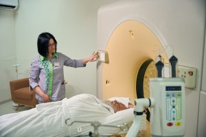
Your High-Field Scan Experience
A high-field MRI scan—such as those using 3T or 7T magnets—provides speed and clarity that’s hard to match. It shifts the focus to patient comfort. These scans provide medical teams with higher quality images, which is crucial for difficult diagnoses or research.
We understand that the process can seem intimidating, particularly if it’s your first time. Here’s a complete overview of your experience from beginning to end! We’ll be particularly interested in hearing about how U.S. Providers have prioritized patient experience and needs.
Simple Steps: Getting Prepared
-
Arrive early to complete any forms.
-
Provide as much detail as possible regarding your medical history, particularly with regard to the presence of any implants, allergies, or previous surgeries.
-
Wear loose, metal-free clothing.
-
Dietary restrictions: Follow dietary restrictions if provided (rare, only for abdominal scans).
-
Remove all metal (jewelry, watches, hairpins) before entering.
Letting your technologist know your health history can prevent safety concerns from arising. Metal can create serious issues in the scanner, so it’s always good to double check before you step in the door.
What Happens Inside the Scanner
Once you’re inside, you will recline on a padded table that slides through the opening of the scanner. Depending on the body part being scanned, the scan takes 15–45 minutes. Remaining motionless during your scan is essential in order to obtain high-quality images.
Typically, short bore MRIs are the most widely used. Their larger, wider and shorter design not only intimidates patients less but addresses anxiety and feelings of claustrophobia. You can expect minor discomfort, but the reduced scan times with high-field systems—pluses in both comfort and convenience—make a big difference.
Understanding Scanner Noises
The scanner produces sudden, loud knocking or tapping noises—these are caused by fast, switching magnetic gradients. Noise-cancelling headphones or earplugs are incredibly helpful. Communicating these concerns to your technologist is the best policy.
U.S. Focus on Patient Comfort
Facilities have started providing blankets, music, and soothing room layouts. Constructive patient feedback has influenced these changes, creating a more comfortable experience with each scan. Most clinics will allow you to tour the scanner and ask questions prior to your test.
Implant Safety: Our Top Priority
Product safety checks are rigorous. Situations such as pacemakers, metal clips or other implants will be checked for by technologists. Each device is thoroughly vetted prior to the MRI compatibility determination.
For instance, some dental fillings are compatible, while legacy pacemakers are not.
Communicating with Your Technologist
We welcome conversations at each stage. Feel free to inquire about how long the scan will take, how loud the machine will be, or what to expect. Your technologist will walk you through the process and address typical questions such as how to indicate you would like to stop the scan.
Technical Hurdles We Are Overcoming
High-field MRI creates crisper images and new, cutting-edge views to see deep inside the body. Yet at the same time, it creates tremendous opportunities. These scanners utilize very powerful magnets—often 7 Tesla or greater! Even small miscalculations or movements can create large artifacts or safety hazards. To use these tools in real clinics, teams in the U.S. Work hard to fix each challenge, step by step.
Taming Pesky Image Artifacts
At high-field MRI, we run into challenges with “artifacts”—that is, strange shapes, shadows, or gaps that can appear in an image. These are the result of factors such as patient head motion, metallic dental implants, or variations in tissue. Researchers are reducing these problems by employing novel scanning techniques.
They use specialized techniques such as the interleaved narrow-band PRESS sequence and shimming gradient coils. To overcome the challenge of motion artifacts, a subset of sites utilize real-time tracking, as well as advanced pulse sequences. This pushes us to make sure that physicians are receiving clean, consistent images.
Managing Radiofrequency Energy Safely
Since high-field MRI uses more radio waves, more heat can accumulate in the body. Teams keep a very close eye on SAR, or specific absorption rate. New RF coil designs and smarter pulse sequences go a long way toward keeping energy levels as low as possible.
In tandem, sensors and software alert staff if levels exceed safe levels. This delicate balance is what keeps patients safe with each and every scan.
Ensuring Perfect Magnet Stability
Ensuring perfect magnet stability is critical. Just a small movement can ruin an image. To avoid this, engineers implement improved cooling, feedback systems, and designs such as the MAGNUS head gradient coil to ensure fields remain stable.
Perfect magnet stability produces sharper images and more precise scientific results.
Unique High-Field Safety Protocols
Staff receive additional training and must undergo an MRI under the same conditions. Facilities implement these rigid safety protocols, including metal screening and 24/7 surveillance. Unique safety protocols were developed for these more powerful scanners, focusing on patient and staff safety at every stage.
Addressing Cost and Accessibility
High-field MRI is more expensive and not all clinics can swing it. Other organizations have grouped together or utilized shared imaging centers to increase availability. The researchers are working to find cheaper coil designs and smarter software so that more patients can enjoy the benefits.
Innovations Pushing MRI Boundaries
Cutting-edge high-field MRI is going beyond traditional boundaries by combining greater field strengths with more advanced hardware and software. Ongoing academic research and practical engineering in the field fuel these developments. They greatly limit our understanding of the body and brain today.
Smarter Coils for Better Signals
RF coil design has undergone a dramatic revolution in high-field MRI. These new coil arrays are able to pick up more signal coming from the body, allowing for sharper images at higher speeds and less noise.
More advanced setups even employ flexible coils that conform to a patient’s anatomy, yielding more comfortable, accurate scans. For instance, phased-array head coils at 7T allow doctors to visualize the hippocampus with 450-micron resolution. What was once an unattainable level of clarity now serves to detect brain abnormalities earlier.
AI Making Images Even Clearer
AI tools are increasingly becoming available to operate alongside MRI machines in order to automatically clean up images and highlight nuances that previously may have been overlooked. AI algorithms filter out the noise, sharpen prominent features, and even flag unusual patterns.
Deep learning models can identify lesions that are otherwise concealed or minor tissue modifications, aiding radiologists in difficult scenarios. For example, AI is able to combine scans from multiple types of MRIs. This unique combination displays both anatomy and blood flow together, providing a more comprehensive view.
Novel Ways to See Inside You
Novel MRI techniques have allowed researchers to interrogate the brain at the cellular level. High-field MRI at 7T is particularly good at differentiating gray and white matter.
It has the potential to map very small brain features, like microscopic cortical columns! Together, these advances allow physicians to monitor disease progression and identify issues sooner. The past few decades have seen an increased focus on the study of medial temporal lobe epilepsy. This novel study reveals important insight into how the disease initially develops and advances.
Next-Gen Magnet Technology Arrives
Today’s high-field MRI magnets are lighter and better shielded, partly due to active shielding. This eliminates the need for miles of iron in hospital walls.
With these new magnets come much stronger, steadier fields, translating into higher signal-to-noise ratios and faster scans. Other new designs employ completely different superconducting materials, allowing them to push field strengths even higher while maintaining greater stability within the systems.
Future of Specialized Contrast Agents
With contrast agents moving in different directions, MRI scans are able to capture minute characteristics within tissues and blood flow. Some are even able to display changes at the cellular level, or label specific proteins in the brain.
That translates to clearer, more focused scans for things like cancer or neurodegeneration, allowing for diseases to be caught at an earlier stage.
Faster Scan Times Emerging
Those faster scan times are mainly a result of clever imaging techniques. Parallel imaging, compressed sensing and more efficient pulse sequences have all increased the rate at which MRI can gather data.
This is a tremendous benefit to patients who have difficulty remaining motionless, and ensures clinics can scan more individuals every day. For instance, with some of the newer techniques, physicians can obtain detailed brain images in a matter of minutes instead of hours.
High-Field MRI: The U.S. Outlook
High-field MRI continues to be an important innovation in American healthcare. It gives physicians high-resolution images, as well as quick scan times. This robust provision has a tangible impact especially in areas such as neurology, oncological, and cardiological care. Currently, systems operating at 3 Tesla or above are becoming increasingly prevalent.
At the same time, the mid-field segment continues to dominate the market share with an impressive 42.7% share, as hospitals look for a compromise between cost and performance. The demand for higher quality imaging is snowballing. The U.S. Market is poised to grow by nearly 6.6% annually over the next 10 years!
Facility Design and Installation
Purchasing the machine is just the first step to getting a high-field MRI up and running. Given the large bore size, hospitals need to consider room dimensions, shielded walls, and cooling systems. The machine’s location within the building is important for patient flow, but also for staff members’ safety.
Renovating existing buildings frequently involves moving structural walls or moving electrical raceways, which can extend timelines. Many hospitals implement modular suites, constructed off-site, to accelerate installations.
The Value for American Hospitals
American hospitals that have adopted high-field MRI are able to realize superior patient outcomes. They benefit from quicker diagnoses, which reduces overall costs. These systems allow hospitals to differentiate themselves by being able to provide state-of-the-art care.
Consider this scenario—most university medical centers are already using 3T MRI to establish a research base and draw the best talent.
FDA Approval: Ensuring Standards
The FDA reviews each new MRI to ensure they are safe and accurate. This process involves extensive laboratory tests, clinical trials, and FDA review to ensure that each machine adheres to rigorous standards.
In just the past few years, the FDA has already approved multiple new coils and software updates, illustrating the rapid pace of this advancing field.
Training Our Imaging Specialists
Keeping a high-field MRI properly tuned and running efficiently requires a high level of expertise. Techs require additional training on the new systems and need to stay constantly updated on emerging tech by obtaining continuing education certificates.
Leading programs, such as the one offered by UCSF or Mayo Clinic, emphasize both in-person practical training and virtual education.
Eco-Friendly System Designs
These days, more makers seek to minimize waste generation and energy consumption. Innovation since newer machines require less helium and utilize more efficient cooling, thereby reducing their carbon footprint.
Many U.S. Hospitals are signing on to environmentally friendly programs that help them monitor and reduce energy consumption.
Integrating into Healthcare Workflows
Adding MRI to hospital routines requires collaboration. Nurses, techs, and doctors need to be able to pass information quickly to provide the best care for patients. Other hospitals have implemented digital scheduling, supported by AI, to accelerate imaging and reduce wait times.
Conclusion
Advanced high-field MRI provides high resolution images and cutting-edge methods to visualize the human body. American physicians are now finding signs of trouble earlier and organizing care more effectively. The technology continues to rapidly improve. Teams troubleshoot previous limitations and devise novel approaches to obtain even higher resolution images. A scan today seems easy and trivial, not like an event. Countless community-led clinics in San Francisco and across the country are responsible for making this equipment a day-to-day reality. Residents experience tangible outcomes that inform and steer major health decisions. Interested in learning how this technology can benefit you or your agency? Follow along by continuing to question your care provider and engage in shared decision-making! Need some more inspiration? Look into communities where actual users and technology professionals discuss their experiences and recommendations.
Frequently Asked Questions
What is an advanced high-field MRI?
What is an advanced high-field MRI? It produces remarkably sharp pictures of your body’s internal structures. As a result, this advanced technology allows physicians to see a much greater area, diagnosis is faster and more precise.
How does high-field MRI improve image quality?
Improved image quality High-field MRIs generate stronger signals, which lead to more detailed, higher-contrast images. This unprecedented level of detail allows physicians to identify problems that would otherwise be overlooked in conventional MRI systems.
Is high-field MRI available in the United States?
Is high-field MRI available in the United States? The technology continues to be adopted in metropolitan areas and rapidly expanding into more suburban and rural communities.
What can I expect during a high-field MRI scan?
You’ll need to lie still while the MRI scanner takes images. In general, the procedure is safe, painless, noninvasive and usually completed in 30–60 min. High-field MRI scans are often much noisier, so you might be provided with ear protection.
Are high-field MRI scans safe?
Yes. Unlike X-rays and CT scans, high-field MRIs do not use radiation, making them extremely safe for most individuals. If you have an implantable device or other metal implant, you should inform your physician prior to receiving an MRI scan.
What makes high-field MRI important for American healthcare?
Advanced high-field MRI allows U.S. Doctors to diagnose diseases earlier and more accurately. As a result, treatment plans are more informed and patient outcomes are enhanced nationwide.
What challenges are being addressed with high-field MRI technology?
U.S. Specialists are working on shortening scan times and increasing patient comfort in the machines, which are loud and intimidating. Additionally, they are working to establish safe usage standards for those with implants. This is where new innovations really make it possible to tackle these challenges.